EMERGENCY: If your animal is in immediate danger or showing signs of distress, contact your veterinarian or nearest emergency animal hospital FIRST. The information below is for educational reference only. Guardians.dog and its representatives do not provide veterinary care.
The Invisible Threat: Ticks, Mosquitoes, and Arthropod Pathogens
Livestock Guardian Dogs live full-time in the pasture alongside the same wildlife that harbors dangerous environmental ticks and mosquitoes. Unlike indoor pets, an LGD cannot easily avoid peak mosquito hours or deep brush. To ensure their longevity, handlers must understand symptoms, diagnosis, and prevention of vector-borne illnesses.
The Co-Infection Threat: Because a working LGD is exposed to thousands of bites over a season, simultaneous infections (such as contracting Babesiosis via ticks while also battling Dirofilariasis from mosquitoes) are highly common. These extreme co-infections compound morbidity, drastically suppress the immune system, and confound veterinary diagnosis.
Interactive USA Risk Map
Want to see the specific tick and mosquito threat levels in your county? Use our interactive map tool to view regional densities for Lyme, Heartworm, and other vector-borne diseases.
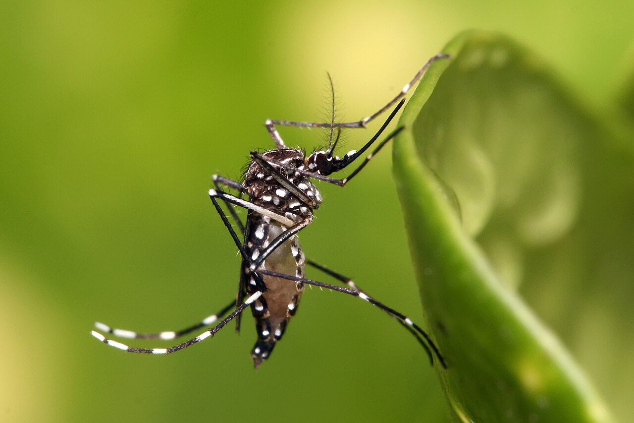
Aedes aegypti (Mosquito vector) Source: CDC / Public Domain
Vector: Mosquitoes (including Aedes aegypti in South Texas)
Heartworm disease is caused by a parasitic worm (*Dirofilaria immitis*) that lives in the heart, lungs, and associated blood vessels of affected dogs, causing severe heart failure, organ damage, and death. Coyotes are a primary wildlife reservoir for heartworm, placing LGDs living in coyote-heavy territories at heightened risk. Recent studies confirm that the notorious Aedes aegypti mosquito is actively driving extreme heartworm prevalence in South Texas.
Symptoms & Progression:
Early Stage: Often asymptomatic or occasional minor cough.
Moderate Stage: Persistent cough, fatigue after moderate activity, exercise intolerance (the dog stops patrolling).
Severe Stage: Weight loss, swollen belly (from fluid accumulation in the abdomen due to heart failure), labored breathing, and coughing up blood.
Vector-borne disease concerns should be evaluated by a veterinarian, who can guide testing, prevention, and management based on the dog’s exposure risk and symptoms.
Diagnosis requires a standard blood test (antigen test). Some vector-borne disease cases may require complex veterinary management, so prevention, early testing, and prompt professional guidance are especially important.
Prevention: No vaccine exists. Must be controlled via monthly systemic preventatives. Since mosquito activity is extending longer into the cold seasons globally, year-round prevention is strongly advised for farm dogs in endemic areas.
Lyme Disease (Lyme Borreliosis)
Lyme Disease
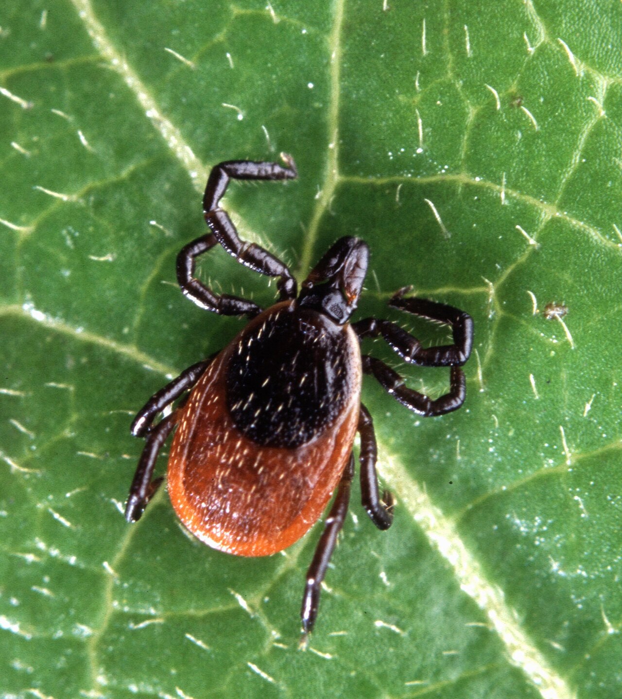
Ixodes scapularis (Deer tick) Source: CDC / Public Domain
Vector: Deer Ticks (Black-legged ticks)
Caused by the *Borrelia burgdorferi* bacteria, Lyme disease is transmitted when an infected tick remains attached and feeds for at least 24-48 hours. LGDs sharing pasture edges with deer are at extremely high constant risk.
Symptoms & Progression:
"Shifting-leg" Lameness: The hallmark sign. The dog may limp on one leg for a few days, then seemingly recover, only to limp on a different leg the next week due to traveling joint inflammation.
Swollen, warm, or painful joints.
Lethargy, fever, and reduced appetite.
Kidney Damage: Severe untreated cases can progress to fatal Lyme nephritis.
Vector-borne disease concerns should be evaluated by a veterinarian, who can guide testing, prevention, and management based on the dog’s exposure risk and symptoms.
Often diagnosed using a SNAP 4Dx blood test. Positive vector-borne disease results should be reviewed with a veterinarian, who can help determine whether monitoring, prevention changes, or medical care are appropriate.
Prevention: Lyme vaccines are available (starting at 8 weeks, with 2 doses and an annual booster). However, since vaccines do not stop the tick from biting and transmitting *other* diseases, high-quality topical or oral tick preventatives are the actual first line of defense.
Ehrlichiosis & Anaplasmosis
Tick-Borne Rickettsial Diseases
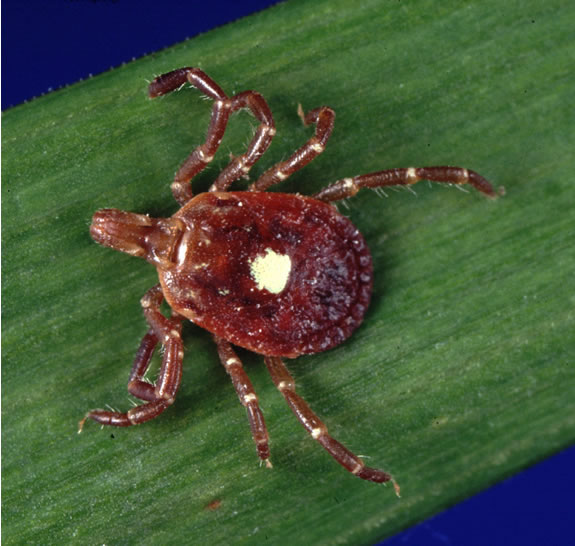
Amblyomma americanum (Lone Star tick) Source: CDC / Public Domain
Vector: Brown Dog Ticks, Deer Ticks, Lone Star Ticks, Mosquitoes, Fleas
These two diseases are caused by similar types of intracellular bacteria (*Ehrlichia spp.* and *Anaplasma spp.*). They invade the dog's white blood cells. While Lyme attacks the joints, these diseases often attack the blood and immune system.
Symptoms & Progression:
Acute Phase: Fever, lethargy, swollen lymph nodes, and spontaneous bleeding (such as unexplained nosebleeds or bruising on the belly from low platelet counts).
Subclinical Phase: The dog may seem totally normal for months or years while the organism hides in the spleen.
Chronic Phase: Severe weight loss, anemia, bleeding disorders, and neurological signs.
Vector-borne disease concerns should be evaluated by a veterinarian, who can guide testing, prevention, and management based on the dog’s exposure risk and symptoms.
Diagnosed via blood testing (frequently on the same SNAP 4Dx test that catches Lyme and Heartworm). Positive vector-borne disease results should be reviewed with a veterinarian, who can help determine whether monitoring, prevention changes, or medical care are appropriate.
Zoonotic / Shared Risk (RMSF Spotlight): While dogs do not transmit rickettsial diseases directly to humans, an infected LGD acts as an environmental sentinel. If your dog contracts Rocky Mountain Spotted Fever (RMSF), it means the pasture environment is heavily loaded with Rickettsia rickettsii-infected ticks, placing you and your family at immediate shared risk.
Zoonotic Vector-Borne Protozoa
Unlike standard bacterial infections, protozoal infections are caused by single-celled organisms that are incredibly difficult to clear once established. For the diseases below, the domestic dog serves as a primary biological reservoir, elevating the zoonotic threat to humans living in the same environment. Dangerously, many infected dogs remain completely asymptomatic, acting as silent carriers that allow local sandflies and kissing bugs to transmit the pathogen to the farm family.
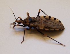
Triatominae (Kissing Bug) Source: CDC / Public Domain
Vector: Triatomine Bugs ("Kissing Bugs")
Caused by the protozoan Trypanosoma cruzi. Dogs are the primary domestic reservoir for this pathogen across the Americas. Kissing bugs feed on the dog and defecate into the bite wound, transmitting the parasite. Chagas causes chronic, irreversible cardiac manifestations (myocarditis, severe arrhythmias, and sudden right-sided heart failure) in dogs.
Sentinel Warning: A Chagas-positive LGD indicates a high density of infected kissing bugs around the farm, representing an extreme risk of lethal Chagas disease for the handlers and their children.
Leishmaniasis ZOONOTIC THREAT
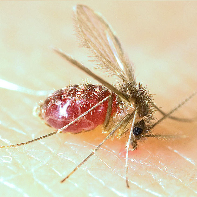
Lutzomyia (Sandfly) Source: Public Domain
Vector: Sandflies (Lutzomyia spp.)
Autochthonous US Spread: Once considered an import-only "foreign" disease, clinical data confirms Leishmaniasis is now actively and natively spreading within the USA (specifically cutaneous forms in Texas) via Lutzomyia sand flies.
Caused by Leishmania infantum (Visceral) and L. mexicana (Cutaneous). LGDs act as the main domestic reservoir. The protozoa live inside the dog's macrophages (white blood cells), causing chronic kidney failure, extreme weight loss, and severe skin lesions (especially around the muzzle and eyes). Because it hides inside host cells, it is essentially incurable—clinical management only suppresses the parasite load.
Babesiosis ZOONOTIC THREAT
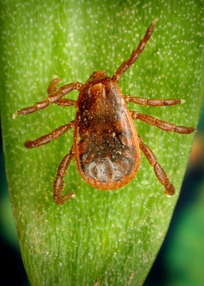
Rhipicephalus sanguineus (Brown Dog Tick) Source: Public Domain
Vector: Ticks (Brown Dog Tick) and Direct Dog Bites (B. gibsoni)
Caused by Babesia species, these protozoa invade and destroy the dog's red blood cells. While typically transmitted by ticks, the B. gibsoni strain is highly transmissible directly through dog bites—making it a severe risk for working LGDs that regularly engage in physical combat with feral dogs or coyotes.
Symptoms & Progression:
Acute Hemolytic Anemia: The hallmark sign. As red blood cells are destroyed, the dog becomes profoundly weak.
Visual Indicators: Pale or severely jaundiced (yellow) gums, and notably dark red or orange "coffee-colored" urine due to hemoglobinuria.
Organ Involvement: Fever, lethargy, and a significantly enlarged spleen (splenomegaly) upon palpation.
Vector-borne disease concerns should be evaluated by a veterinarian, who can guide testing, prevention, and management based on the dog’s exposure risk and symptoms.
Diagnosis is confirmed via blood smear or PCR testing. Care decisions for vector-borne disease depend on the organism involved, test results, symptoms, and the dog’s overall health history:
For B. canis: Intramuscular injections of Imidocarb dipropionate, often pre-treated with atropine to manage harsh cholinergic side effects.
For B. gibsoni: An intensive oral protocol combining atovaquone (given with a fatty meal for absorption) and azithromycin for 10 days.
Zoonotic Warning: Babesiosis presents a life-threatening risk to human handlers who are immunocompromised or have had a splenectomy.
Bartonellosis & The Occupational Hazard
Bartonellosis ZOONOTIC EMERGENCY
Ctenocephalides felis (Primary Flea Vector) Source: Public Domain
Vector: Fleas, Ticks, and Direct Scratch/Bite Transmission
Bartonella species are stealth intracellular bacteria. While classic transmission occurs via fleas and ticks, Bartonella is highly transmissible via scratches or bites if flea dirt is present in the dog's nail beds or saliva.
Symptoms & Progression:
Asymptomatic Stealth: Many LGDs remain subclinical carriers for years, acting as silent environmental reservoirs.
Cardiac Failure: When clinical, it famously attacks the heart valves, causing severe endocarditis, new heart murmurs, and congestive heart failure.
Systemic Inflammation: Unexplained lameness/arthritis, swollen lymph nodes, and recurrent fevers.
Vector-borne disease concerns should be evaluated by a veterinarian, who can guide testing, prevention, and management based on the dog’s exposure risk and symptoms.
Diagnosis is notoriously difficult (often requiring advanced PCR or BAPGM enrichment culture) because the bacteria hide inside blood vessel walls.
Long-Term Antibiotics: Eradicating the bacteria is extremely difficult. It requires an aggressive, multi-week (4-6+ weeks) combination therapy.
Protocol: Veterinarians frequently use a combination of doxycycline alongside a fluoroquinolone like enrofloxacin to penetrate the intracellular spaces.
The Handler Warning: Clinical literature documents severe cases of veterinary workers and dog handlers contracting tripartite co-infections (Anaplasma, Bartonella, and Mycoplasma) directly from constant, close-proximity work with domestic dogs. Handlers removing ticks/fleas without gloves or suffering accidental bites/scratches during field work are at direct, life-altering risk.
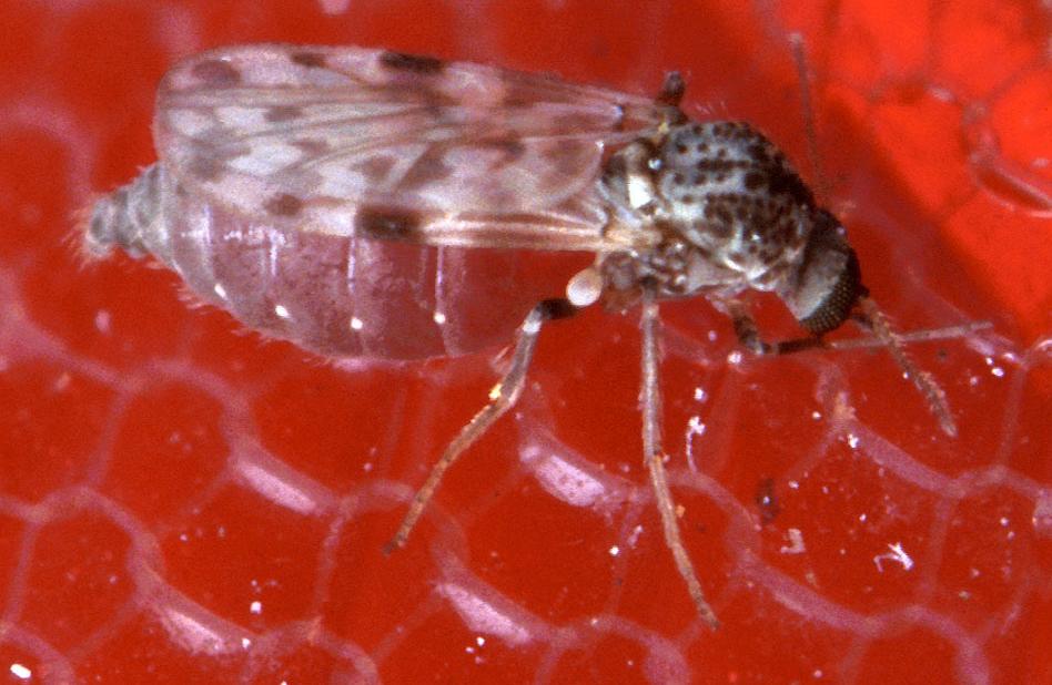
Ceratopogonidae (Biting Midge) Source: Public Domain
Vector: Biting Midges (*Culicoides*) and Black Flies (*Simulium*)
Historically restricted to the Mediterranean basin, modern disease mapping confirms that these filarial worms are now successfully leveraging North American midges and black flies to infect dogs across the American Southwest (California, Arizona, New Mexico). Adult worms form acute, painful nodules on the dog's eyes (sclera/conjunctiva), potentially blinding the LGD, and the zoonotic spillover to humans causes similar ocular trauma.
Flea-Borne Bacterial Vectors
Plague (*Yersinia pestis*) ZOONOTIC THREAT
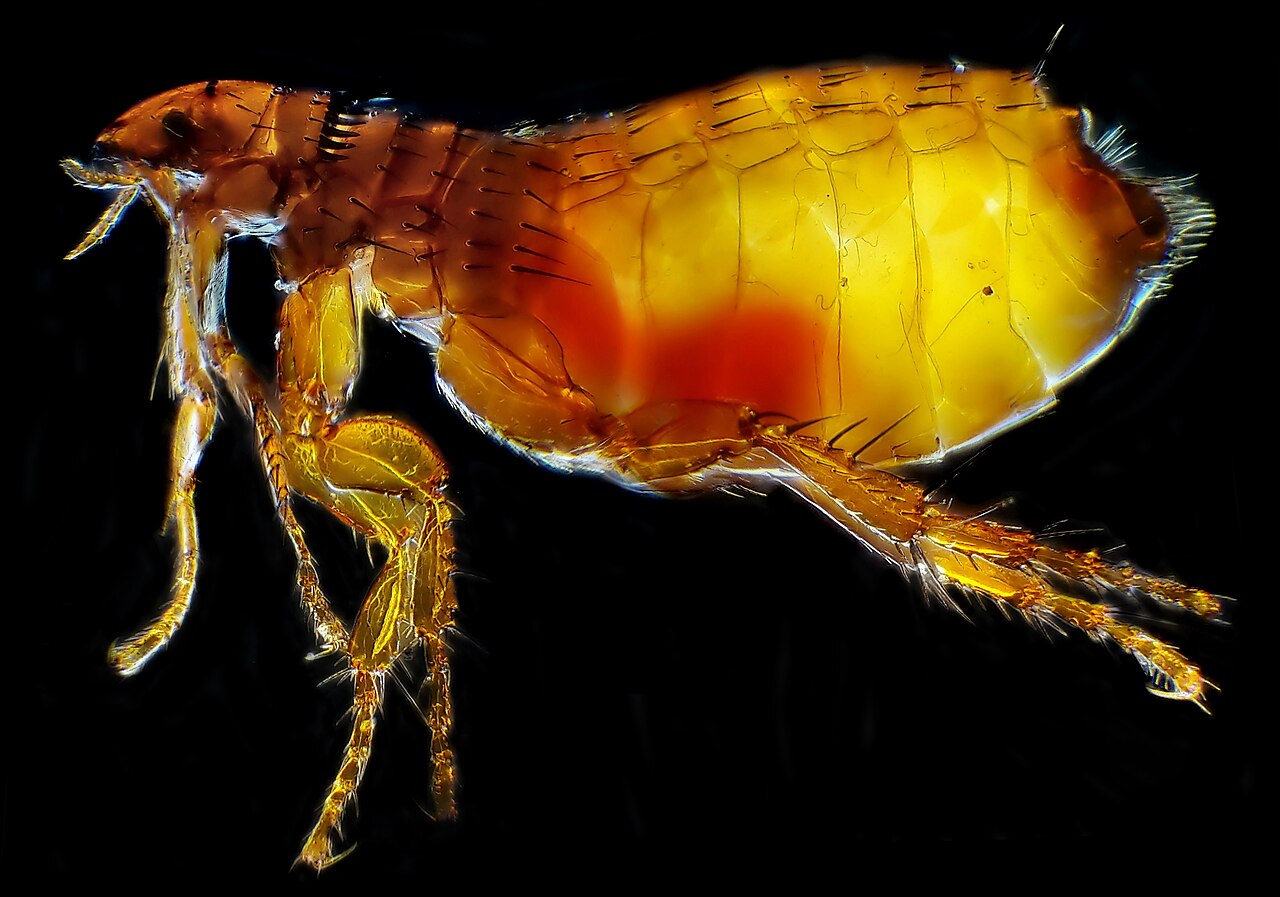
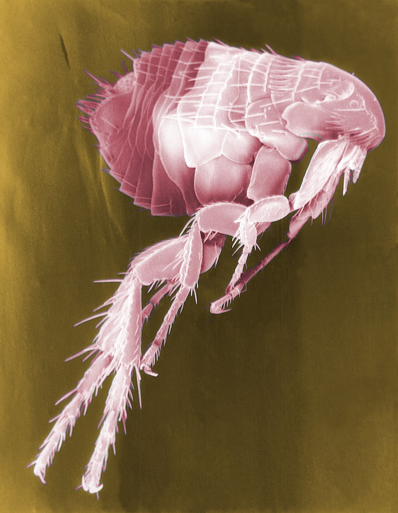
Flea Vector (Scanning Electron Micrograph) Source: CDC / Public Domain
Vector: Fleas (specifically from Prairie Dogs, ground squirrels, and rodents)
In the American West and Southwest, rural LGDs frequently hunt and kill prairie dogs to protect their pastures. Prairie dog colonies are massive reservoirs for the Yersinia pestis bacterium—the exact same pathogen responsible for the Black Death. When the LGD kills the prairie dog, infected fleas quickly abandon the dead host and jump onto the dog.
The Zoonotic Danger: While dogs are somewhat more resistant to the Plague than cats, they can develop bubonic plague (severely swollen lymph nodes) or pneumonic plague (severe coughing). The extreme danger is that the LGD acts as an active vector, bringing the infected prairie dog fleas directly back to the handler, or transmitting pneumonic plague directly to humans via aerosolized respiratory coughing.
Regional / Global Diseases
African Canine Trypanosomiasis
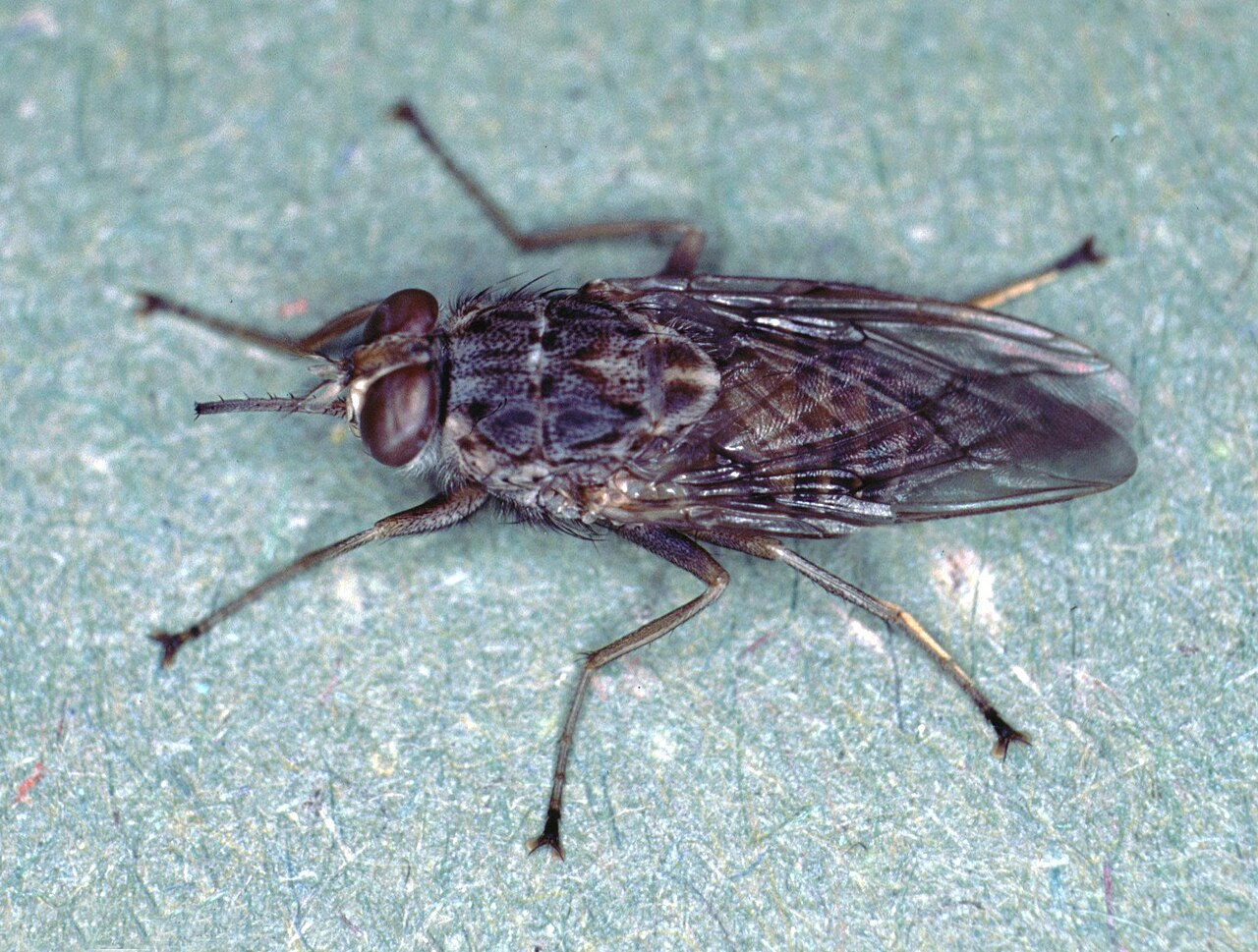
Glossina (Tsetse Fly) Source: Public Domain
Vector: Tsetse Fly
For working LGDs utilized in Southern and Eastern African conservancies (such as anti-poaching or cheetah-conservation LGDs), the Tsetse fly poses a severe threat. There is currently no proven preventative vaccine. Handlers must rely on fly-proof kenneling during peak activity hours and altering grazing routes to avoid dense fly infestations.
Advanced Prevention Strategy (One Health)
Isoxazolines & The Wildlife-Livestock Interface
Working dogs do not live in a vacuum—they live on the pasture with livestock. While LGDs protect the herd from predators, they are surrounded by Culicoides biting midges that transmit devastating agricultural diseases like Bluetongue Virus and Epizootic Hemorrhagic Disease (EHD) to the sheep/cattle.
Clinical entomology demonstrates that Isoxazoline-class preventatives (e.g., fluralaner / Bravecto) actively kill adult biting midges that feed on the dog. By treating your LGDs with systemic isoxazolines, the dog essentially acts as an active, localized insecticidal net—slaughtering the midge populations around the herd while simultaneously protecting the dog from Lyme and Onchocerca eyeworms. This is a foundational standard of modern One Health biosecurity.
Bowman, D. D., et al. (2009).Heartworm disease in dogs: a review. Veterinary Parasitology.
Breitschwerdt, E. B. (2014).Bartonellosis: One Health Perspectives for an Emerging Infectious Disease. ILAR Journal.
Otranto, D., et al. (2018).Efficacy of fluralaner (Bravecto) against Culicoides biting midges. Parasites & Vectors.
Petersen, C. A., & Barr, S. C. (2009).Canine leishmaniasis in North America. Veterinary Clinics of North America: Small Animal Practice.
CDC / NIOSH. (2020).Tick-Borne Diseases - Occupational Hazards and Prevention. National Institute for Occupational Safety and Health.
About the Author
Written by Jesika VanFossen — LGD breed consultant with 28 years of hands-on livestock guardian dog experience, 30+ years of animal husbandry, and founder of URMAH LC, the longest-running registered Turkish Boz Shepherd breeding program in the United States. Trained by Turkish dog experts, Jesika directs a network of breed-specific research platforms including Guardians.dog, Database.dog, and TurkishBoz.com, and maintains an active network of procurement specialists, along with veterinary, behavioral, and breed professionals for expert referral.
Educational Only: This information is general guidance based on current research and typical breed characteristics. Individual dogs and situations vary significantly. Always consult qualified professionals for specific advice.
Disclaimer: Health & Veterinary Disclaimer: This content is for general educational purposes only and is not veterinary, medical, diagnostic, treatment, dosage, or emergency advice. Dog health needs vary by individual animal, breed, age, condition, medication history, pregnancy status, environment, and veterinary care. Always consult a licensed veterinarian or qualified animal health professional before making health, medication, nutrition, breeding, or emergency-care decisions.
Health & Veterinary Disclaimer: This page is for educational purposes only and is not a substitute for veterinary care. LGD breeds and giant-breed dogs may have special health risks, medication sensitivities, and emergency-care needs. This content does not diagnose, treat, prescribe, or replace guidance from a licensed veterinarian. If your dog may be sick, injured, poisoned, or in distress, contact a veterinarian, emergency clinic, or poison-control resource right away.