Allergies are one of the most common chronic health issues in large and giant breed dogs. For livestock guardian dogs, the problem is compounded by constant outdoor exposure to environmental triggers that indoor pets rarely encounter: pasture grasses, hay dust, livestock feed, and agricultural chemicals. Understanding the three types of canine allergies — and how they present differently in working dogs — is essential for keeping your LGDs comfortable and effective.
Key Takeaways
- Gene-Environment Interaction: Canine atopic dermatitis (CAD) is highly heritable (~50% genetic variation), but studies show early rural exposure (farm life) can actually be protective (reducing risk by ~26%) against developing allergies.
- The "Outside-In" Hypothesis: Allergies often begin with a defective skin barrier (abnormal ceramide content, low filaggrin) that allows environmental allergens like dust mites and pollens to penetrate the skin and trigger epicutaneous sensitization.
- Microbial Amplification: Over 54–72% of atopic dogs develop secondary bacterial (pyoderma) or yeast infections, which significantly amplify the inflammatory itch-scratch cycle.
The Three Types of Canine Allergies
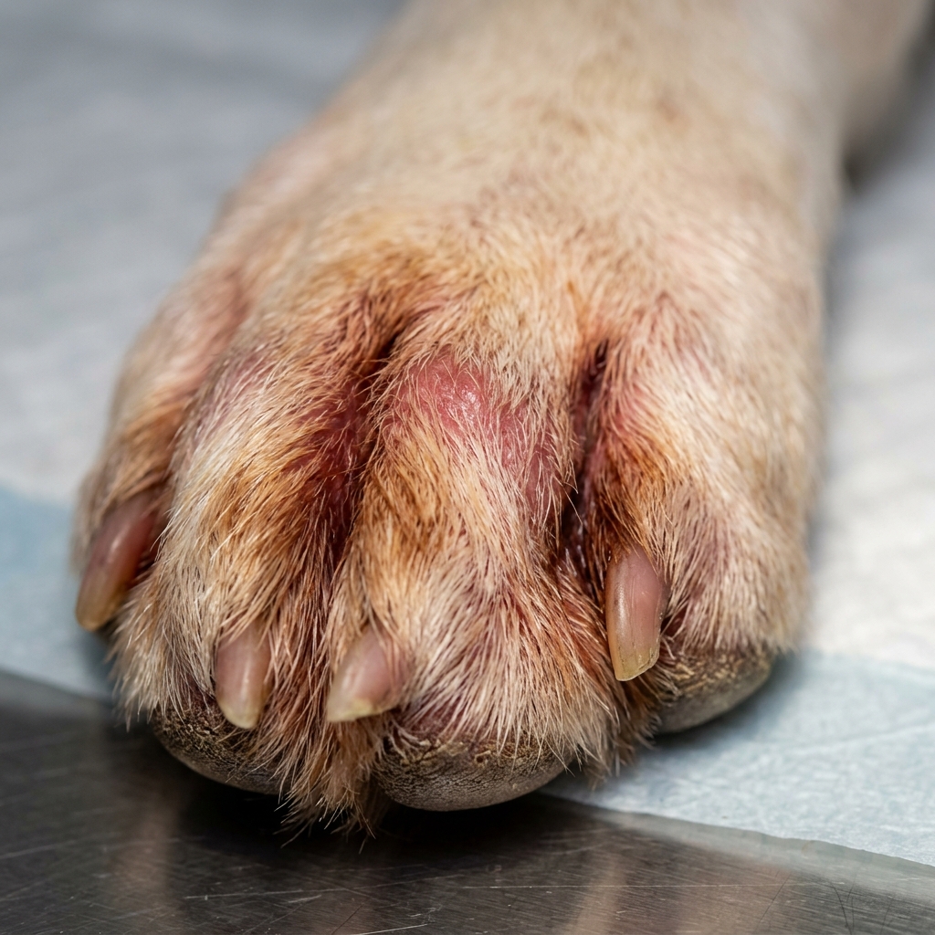
Farm-Specific Triggers
LGDs encounter allergy triggers that most pet dogs never face. Understanding these farm-specific irritants helps you identify and reduce exposure:
Common Farm Allergens for LGDs
- Pasture grasses & weeds: Timothy, orchard grass, Bermuda grass, ragweed, and other pasture species are potent pollen producers. Your LGDs walk through, lie in, and breathe these all day.
- Hay & straw dust: Hay storage areas release fine particulate matter that irritates airways and skin. Mold spores in improperly cured hay are a secondary trigger.
- Livestock feed: Grain dust from cattle, sheep, or poultry feed. Soy-based feeds are particularly allergenic.
- Manure & bedding: Ammonia from urine-soaked bedding irritates skin and respiratory tissue. Cedar chips (sometimes used in dog houses) contain aromatic oils that cause contact dermatitis.
- Agricultural chemicals: Herbicides, pesticides, pour-on livestock treatments, and fly sprays. Even residue on treated pasture can cause contact reactions on paws and belly.
- Insect bites: Flea allergy dermatitis (FAD) is technically an allergy, not just an infestation. Some dogs have extreme reactions to a single flea bite. See our External Parasites guide.
Environmental vs. Food: How to Tell the Difference
| Characteristic | Environmental (Atopic) | Food Allergy |
|---|---|---|
| Seasonality | Seasonal (spring–fall) or perennial | Year-round, no seasonal pattern |
| Age of onset | Usually 1–3 years | Any age; often <1 or >6 years |
| GI symptoms | Rare | Common (soft stool, gas, vomiting) |
| Ear infections | Common | Very common (often the primary sign) |
| Response to steroids | Good (reduces itch quickly) | Partial or poor |
| Definitive test | Intradermal skin testing | 8–12 week elimination diet trial |
Diagnostic Approaches
For Environmental Allergies
- Intradermal skin testing (IDST): The gold standard. A veterinary dermatologist injects tiny amounts of common allergens under the skin and observes reactions. This identifies specific triggers for immunotherapy.
- Serum allergy testing: A blood test that measures IgE antibodies to specific allergens. Less accurate than IDST but more accessible in rural areas. Useful as a starting point.
For Food Allergies
Weeks 1–2: Select a Novel Protein Diet
Choose a protein and carbohydrate source your dog has never eaten before. Common options: venison + sweet potato, rabbit + pea, or a veterinary hydrolyzed protein diet. Feed ONLY this. Avoid table scraps, flavored medications, and new food items unless approved by your veterinarian.
Weeks 2–6: Strict Adherence
The hardest phase for farm dogs. Your LGD cannot eat livestock feed, chew on carcasses, or receive any food outside the trial diet. This may require temporary management changes. One “cheat” resets the clock.
Weeks 6–8: Evaluate Improvement
If symptoms improve significantly (50%+ reduction in itching, ear infections, GI signs), food allergy is likely. If no improvement, food allergy is ruled out — focus shifts to environmental causes.
Weeks 8–12: Challenge Phase
Reintroduce original food ingredients one at a time, every 1–2 weeks. When symptoms return, you have identified the specific trigger protein. This information guides long-term diet selection.
Management Strategies for Working LGDs
Managing allergies in a dog that lives and works outdoors 24/7 requires practical, farm-adapted strategies:
Environmental Allergy Management
- Paw rinses: After heavy pollen days, rinse your dog’s paws with cool water to remove allergen residue. A shallow foot-bath tray near the dog house works well.
- Clean bedding areas: Wash or replace bedding in dog houses weekly during allergy season. Avoid straw bedding (harbors mold) — use washable blankets or elevated cot-style beds.
- Omega-3 fatty acids: Fish oil supplementation (EPA/DHA) improves skin barrier function and reduces inflammatory response. Omega-3 supplementation should only be used at an amount recommended by a veterinarian, especially for large breeds, pregnant dogs, dogs on medications, or dogs with medical conditions.
- Medicated baths: Chlorhexidine or oatmeal-based shampoos every 1–2 weeks during flare-ups. Bathing removes surface allergens and soothes irritated skin.
- Immunotherapy (allergy shots): For dogs with confirmed environmental allergies via IDST, Allergy testing and immunotherapy should be discussed with a veterinarian, especially when symptoms are ongoing or severe. It requires 6–12 months to take effect but can provide long-term relief. Available as injections or sublingual (oral) drops.
Medication Options
- Apoquel (oclacitinib): Fast-acting itch relief. Works within 4–24 hours. Often tolerated for long-term use when prescribed by a veterinarian. Requires veterinary prescription and periodic blood monitoring.
- Cytopoint (lokivetmab): Injectable antibody that blocks itch signaling for 4–8 weeks per injection. No daily pills — well-suited for working dogs that are hard to medicate daily.
- Antihistamines: Over-the-counter options (cetirizine, diphenhydramine) help some dogs. Less effective than Apoquel/Cytopoint but safe and inexpensive. Consult your vet for dosing.
- Steroids (prednisone): Powerful anti-inflammatory for acute flare-ups but not recommended for long-term use due to side effects (increased thirst/urination, immune suppression, weight gain). Use as a rescue therapy, not maintenance.
Research Briefing: Genetics and Barrier Dysfunction
Recent studies in veterinary dermatology frame canine allergic skin disease not just as an immune overreaction, but as a complex convergence of genetic susceptibility, epidermal barrier dysfunction, and microbial dysbiosis.
I. Genetic Load and Early Life Exposure
Atopic dermatitis is highly heritable. In large breeds like Retrievers and German Shepherds, researchers estimate that approximately 50% of the variability in pathogenesis is inherited, while 50% is environmental. For example, a specific risk haplotype in German Shepherd Dogs confers a massive 5.3x increased odds of developing CAD. However, environment matters heavily: studies show that dogs born in rural locations and exposed to natural environments early in life have a significantly reduced risk (OR 0.74 and 0.48, respectively) of developing CAD.
II. The "Outside-In" Epidermal Defect
Mounting evidence points to a primary defect in the skin's barrier function (the "outside-in" hypothesis). Atopic dogs often exhibit abnormal stratum corneum lipid lamellae, reduced ceramide content, and diminished filaggrin expression. This impaired barrier allows environmental allergens (like house dust mites, which are the most common trigger) to penetrate the skin easily. This epicutaneous exposure sensitizes the immune system, leading to a Th2-polarized inflammatory response. Once established, scratching and inflammation cause further barrier damage in a "vicious cycle."
III. Microbial Amplification
Skin barrier dysfunction and cutaneous microbial dysbiosis are now considered essential players in CAD pathogenesis. Clinical datasets show that 54% to 72% of atopic patients suffer from secondary pyoderma (bacterial infection), and over 30% develop Malassezia (yeast) dermatitis. These microbes are not just incidental; they actively perpetuate and amplify the allergic immunological response.
When to See a Veterinary Dermatologist
Consider referral to a board-certified veterinary dermatologist (DACVD) if:
- Your dog has more than 3 ear infections per year
- Symptoms do not respond to standard treatments (Apoquel, Cytopoint)
- You want to pursue immunotherapy (allergy shots) — IDST requires specialist equipment
- Your dog has concurrent skin infections (bacterial pyoderma, yeast dermatitis) that keep recurring
- You suspect a food allergy but elimination diets are impractical for your farm setup
Bizikova, P., et al. (2015). Review: Clinical and histological manifestations of canine atopic dermatitis. Veterinary Dermatology, 26(2), 79–e24.
Cosgrove, S. B., et al. (2015). Efficacy and safety of oclacitinib for the control of pruritus and associated skin lesions in dogs with canine allergic dermatitis. Veterinary Dermatology, 26(3), 127–e33.
Hensel, P., et al. (2015). Canine atopic dermatitis: Detailed guidelines for diagnosis and allergen identification. BMC Veterinary Research, 11, 196.
Hillier, A., & Griffin, C. E. (2001). The ACVD task force on canine atopic dermatitis: Incidence and prevalence. Veterinary Immunology and Immunopathology, 81(3–4), 147–151.
Mueller, R. S., & Olivry, T. (2017). Critically appraised topic on adverse food reactions of companion animals: Common food allergen sources in dogs and cats. BMC Veterinary Research, 13, 9.
Mueller, R. S., et al. (2016). Critically appraised topic on adverse food reactions: Diagnostic testing. BMC Veterinary Research, 12, 275.
Olivry, T., et al. (2015). Canine atopic dermatitis guidance should be reviewed with a veterinarian, especially when symptoms are ongoing, severe, or recurrent. 2015 updated guidelines from the International Committee on Allergic Diseases of Animals (ICADA). BMC Veterinary Research, 11, 210.
Olivry, T., & Mueller, R. S. (2019). Critically appraised topic on adverse food reactions: Prevalence of cutaneous adverse food reactions in dogs and cats. BMC Veterinary Research, 15, 269.
Santoro, D. (2019). Therapies in canine atopic dermatitis: An update. Veterinary Clinics of North America: Small Animal Practice, 49(1), 9–26.